![]()
 |
||
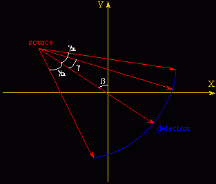
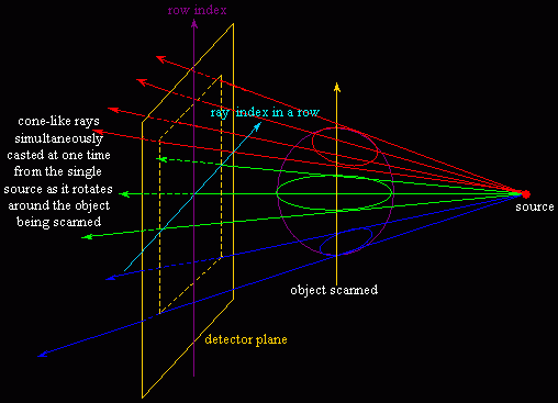
| Computerized Tomography (CT) has been widely used in hospitals and clinics. As a set of rays are cast from a rotating CT scanner to pass through tissues, bones, and whatever organs, the source radiological intensities attenuate to what are measured at the exit by an arrray of detectors to produce projection data, from which a Filtered Back-Projection method based on Fourier Slice Theorem is typically adopted to reconstruct, in frequency domain, a stack of slice images. The patient needs to hold his / her breath when being scanned. Otherwise an incorrect projection dataset would result from shape varying or position changing of human organs as the source (intensity emitter) rotates to complete a 360-degree / 180-degree (full scan / half scan, click to find out how CT rays contribute to image reconstruction) data collection cycle. To relieve the patient from lengthy discomfort caused by the naive Single-Slice Fan Beam Scan (Figure 1), many advanced CT scan modes such as Cone Beam Scan (Figure 2) and Helical Scan and sophisticated reconstruction algorithms have been developed to accelerate the scanning process while maintaining high-quality image generation. | ||
 |
 |
|
|
Figure 1. Single-slice fan beam
scan in the equi-angular case (©
Zhanping Liu).
|
Figure 2. Cone beam scan enabling
multi-slice acquisition (©
Zhanping Liu).
|
|
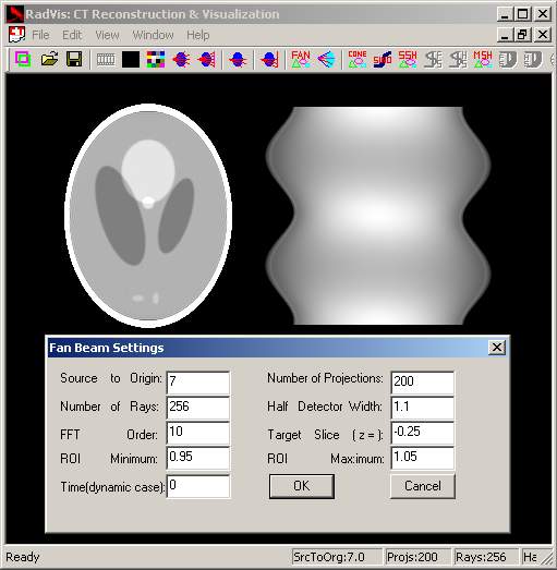
| One problem with the aforementioned approaches is the restriction of their use to stationary objects. How to properly reconstruct motion objects (e.g., lungs) with high temporal-spatial resolution using these scan modalities is still an open problem. Image and Model Based Analysis of Lung Disease (2000.8~2001.5), sponsored by US National Institute of Health (R01 HL64368), was intended to obtain a sequence of high-fidelity images of motion lungs from Multi-Slice Spiral CT data. Thus an important task was to design a sophisticated model (in that physiological knowledge is required) to address the lung motion to guide image reconstruction. A cyliner-based dynamic lung model was devised in place of the traditional ellipsoidal Phantom model to test a variety of CT reconstruction algorithms. RadVis (Figure 3), a simulation system for CT reconstruction and volume data visualization, was developed on Windows NT by Zhanping Liu at the University of Iowa to support Equi-Angular Fan Beam Full / Half Scan, Equi-Spatial Fan Beam Full / Half Scan, Cone Beam Scan, Single-Slice Helical Full / Half Scan, Multi-Slice Helical Full / Half Scan, cylinder-based dynamic lung modeling and reconstruction, volume rendering, and animation production. | ||
 |
 |
 |
|
(a) Fan Beam Scan settings.
|
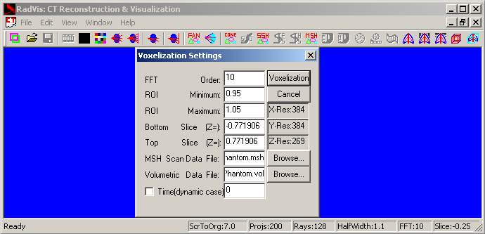
(b) Voxelization to generate a
volume data.
|
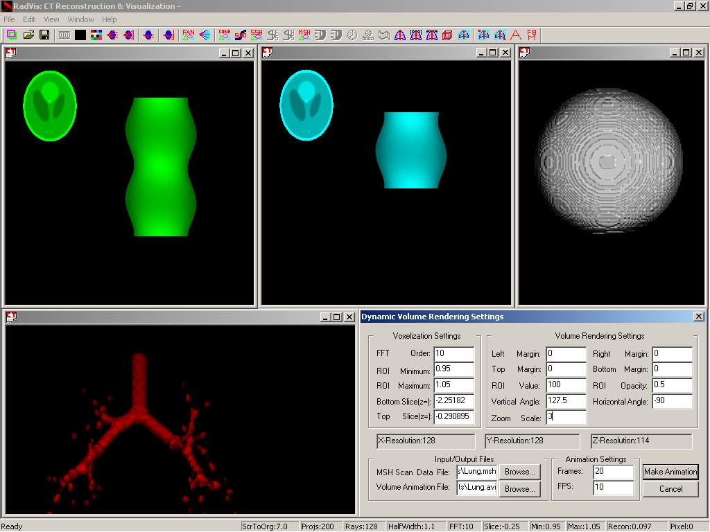
(c) Multi-view RadVis.
|
|
Figure 3. RadVis is a simulation
system for CT reconstruction and volume data visualization.
|